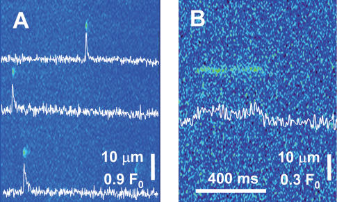
Figure 1. Events in the side-pool regions of fibres under voltage clamp.
Fluo-4 fluorescence in line scans parallel to the fibre axis. Intensity normalized to resting fluorescence (see Methods). A, representative image showing multiple sparks. (Identifier: fibre 072602B record 1.) B, example of a ‘lone ember’. (102102A111.)

