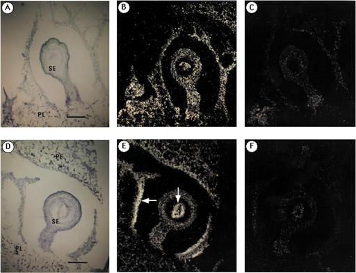
Figure 4.
In situ localization of fructokinase mRNA in seeds and surroundings of very young fruit. Fruit of approximately 10 mm in diameter of wild-type tomato was used for the in situ hybridization. Sections hybridized with Frk1 and Frk2 probes were exposed for 21 and 10 d, respectively. A and D, Bright-field microscopy of the sections hybridized with each probe and stained with KI/I2 and toluidine blue. B, Antisense probe of Frk1 was used. C, Sense probe of Frk1 was used to show hybridization background. E, Antisense probe of Frk2 was used. White arrows indicate the concentrated signals of transcripts. F, Sense probe of Frk2 was used to show hybridization background. Bars represent 200 μm. SE, Seed; PE, pericarp; and PL, placenta.

