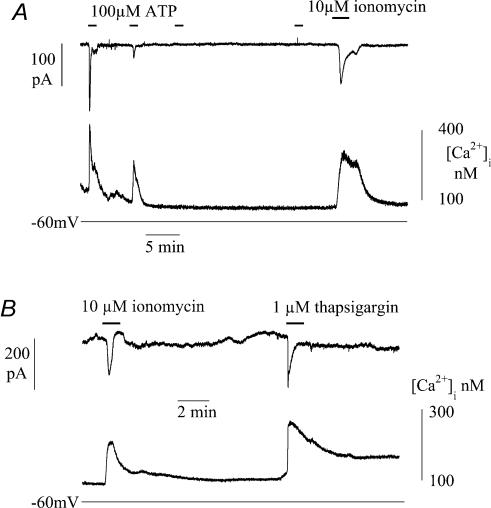
Figure 6. Intracellular pathways for Ca2+ release and uptake in suburothelial myofibroblasts.
A, Ca2+ transients and inward currents during infusion of intracellular low molecular mass heparin by four successive exposures to 100 μm ATP (indicated by horizontal bars). After the last ATP exposure the cell was superfsed with 10 μm ionomycin and the effect of 10 μm ionomycin. B, the effects of 10 μm ionomycin and 1 μm thapsigargin. Cells voltage-clamped with Cs+-filled pipettes at −60 mV.

