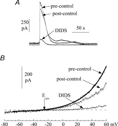
Figure 9. The action of DIDS (1.2 mm) on spontaneous currents in suburothelial myofibroblasts.
A, three superimposed transients from a continuous recording. B, current–voltage relationship of membrane current during a quiescent phase. Membrane potential was changed as a ramp (0.16 V s−1). The line through the pre-control data was best-fitted using eqn (1) (Methods) and the reversal potential, Erev, indicated. Cells were voltage-clamped at 0 mV with a Cs+-filled pipette.

