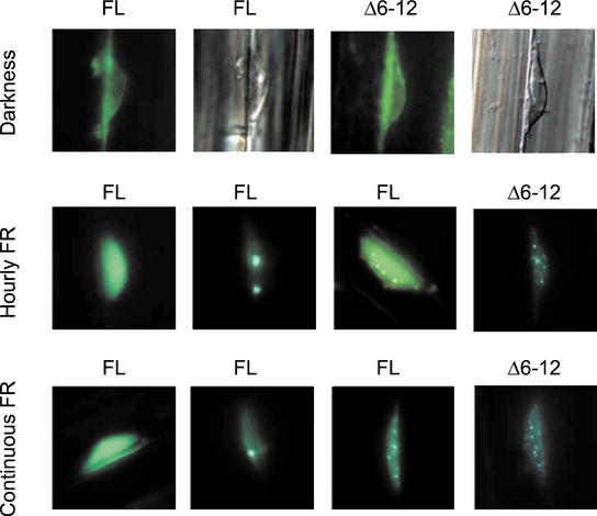
Figure 8.
FL phyA:GFP and Δ6-12 phyA:GFP fusion proteins translocate to the nucleus but show different patterns of nuclear speckle formation under FR. Epifluorescence images of hypocotyl cells from 3- to 4-d-old seedlings of Arabidopsis bearing the oat FL PHYA:GFP or the oat Δ6-12 PHYA:GFP transgene, nonirradiated or exposed to 24 h of either hourly 3-min light pulses of FR (24 μmol m−2 s−1) or continuous FR (1.2 μmol m−2 s−1). The second and fourth pictures of the first row correspond to nuclei that were stained with 4′,6-diamidino-2-phenylindole dihydrochloride to visualize DNA. For both FR conditions, FL oat phyA:GPF transgenics showed (left to right) nuclei with intense background and no spots (the least frequent pattern), nuclei with few small spots, and nuclei with many tiny spots. Δ6-12 oat phyA:GFP fusions only formed numerous tiny nuclear spots.

