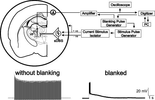
Figure 1. Experimental methods.
Top: schematic representation of sharp intracellular recording set-up in thalamic rat brain slice. Note the two methods of stimulation using either the intranuclear bipolar electrode or the surrounding ‘monopolar-ring’ configuration. Recording and stimulation sites were in ventral-lateral (VL) or ventral-posterior (VP) nuclei. Bottom: intracellular recordings from the same cell with, and without the blanking operation activated.

