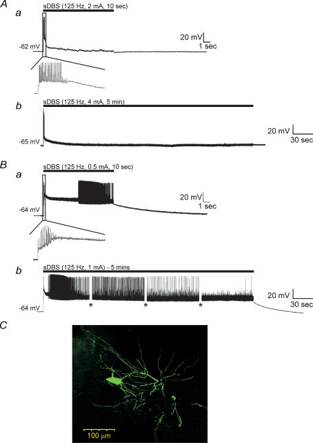
Figure 2. sDBS evoked two distinct types of membrane responses in ventral thalamic neurones.
A, type 1 responses had a large initial depolarization declining toward a smaller but sustained level of depolarization in response to 10 s (a) or 5 min (b) of sDBS. The black bar indicates stimulus onset and duration. B, type 2 responses have a large initial depolarization, which persisted over 10 s (a) or 5 min (b) and led to varied spike activity. The insets show expanded initial responses during the 10 s sDBS train. The amplitude of action potentials in this and following figures were partially reduced due to digitization and some were also truncated or flattened by ‘blanking pulses’. Gaps in the recording (shown as *) indicate times at which current pulse protocols were run during the 5 min sDBS train. C, morphology of a representative ventral thalamic neurone filled with neurobiotin. Note the extensive dendritic tree with numerous distal arborizations.

