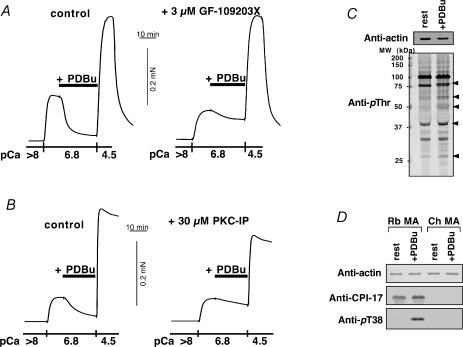
Figure 6. Effect of PKC inhibitors on PDBu-induced relaxation and immunoblots using anti-phosphoprotein antibodies in chicken mesenteric artery.
A, effect of 3 μm GF-109203X on 1 μm PDBu-induced relaxation in α-toxin-permeabilized chicken mesenteric artery (n = 3). Left trace, control PDBu-induced relaxation. Right trace, 3 μm GF-109203X was added 10 min before and present throughout the experiment. B, effect of 30 μm PKC pseudosubstrate inhibitor peptide(19–31) (PKC-IP; the peptide of 2000 Da cannot penetrate cells through α-toxin pores but can through β-escin pores; Kobayashi et al. 1989) on 1 μm PDBu-induced relaxation in β-escin-permeabilized chicken artery (n = 3). Left, control. Right, 30 μm PKC-IP was added 10 min before increase in Ca2+ and was present throughout the experiment. C, protein phosphorylation by PDBu in chicken artery (n = 3). Intact chicken artery strips were exposed to either the normal external solution (rest) or 1 μm PDBu-containing solution for 10 min and quickly frozen. The SDS-extracts were used to monitor phosphorylation levels by immunoblotting using anti-phosphothreonine (anti-pThr) antibody. For immunoblotting for actin, the extracts were diluted 30 times. Arrowheads show more than twice increase in blot density. D, phosphorylation of CPI-17 at Thr38 by 1 μm PDBu in intact rabbit (Rb) and chicken (Ch) mesenteric artery (MA) (n = 3). Top panel shows immunoblots using anti-actin, middle panel using anti-CPI-17, and lowest panel using anti-pThr38 CPI-17 antibodies. SDS-extract containing 20 μg of total protein was applied in each lane for anti-CPI-17 and anti-pT38 immunoblottings. For actin, the extracts were diluted 40 times. Phosphorylation level of CPI-17 at Thr38 was dramatically increased in the PDBu-treated rabbit MA (13, 16). Either polyclonal anti-CPI-17 or anti-pThr38 antibody did not show any bands in chicken MA extracts in the presence and absence of PDBu.

