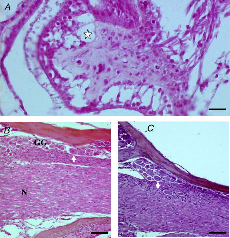
Figure 1. Histology of the IsK−/− vestibular organs.
A, crista of the semicircular canal in an IsK−/− mouse. Note the massive degeneration of the hair cells and the vacuoles in the core of the crista (star). The vestibular ganglion and vestibular nerve in the same IsK−/− mouse (B) and in a wild-type mouse (C). The intact nerve fibres (N) course towards the cristae and the maculae to make contact with hair cells. The soma of the first-order vestibular neurones (arrows), illustrated as a column of cells on the top of B and C, are intact. Scale bars, 30 μm.

