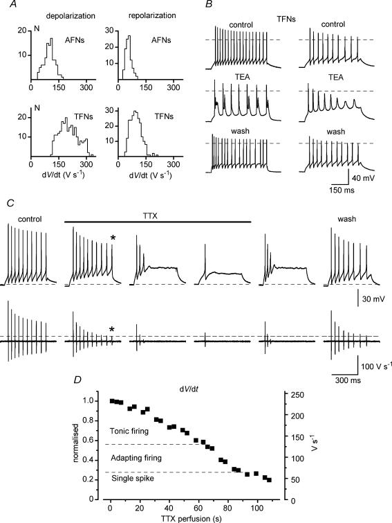
Figure 5. TTX but not TEA can induce adaptation in tonic-firing neurones (TFNs).
A, histograms of distribution of de- and repolarization rates in AFNs and TFNs. Bin width, 10 V s−1. B, modifications of discharge patterns in TFNs by 1 mm TEA. C, induction of adaptation in a TFN during perfusion of the slice with 40 nm TTX. Bottom traces are derivatives of voltage traces (dV/dt). The baselines are 0 V s−1. Dashed line shows the maximum depolarization rate for the reduced spike with the overshoot close to 0 mV (indicated by asterisks). D, modification of firing pattern from tonic to adapting as a function of Na+ channel block during slice perfusion with 40 nm TTX. The maximum depolarization rate of the first spike in a train (right axis) is also shown as normalized to the control value (left axis).

