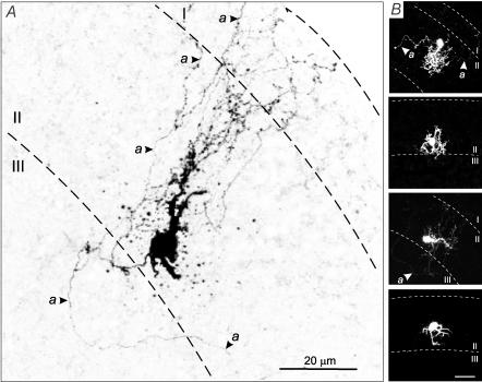
Figure 6. Confocal images of AFNs labelled with biocytin.
A, inverted image of AFN with extensive axonal collaterals indicated by arrows. B, images of another four AFNs two of which have extensive axonal collaterals. Calibration bar, 20 μm. Dashed lines represent the borders between laminae I, II (SG) and III.

