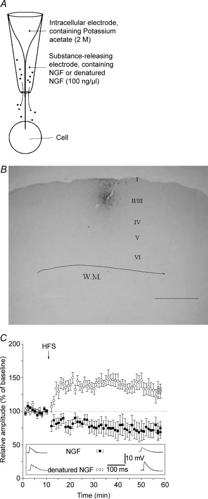
Figure 2. NGF depresses LTP and generates LTD after high frequency stimulation.
A, concentric electrode used to record from the cell and to locally deliver NGF (or denatured NGF). The inner portion is the sharp electrode used for electrophysiological recording. The outer electrode has an opening of about 50 μm and contains the drug to be delivered. B, neurones labelled by uptake of 0.5% biocytin released from the outer electrode (scale bar = 800 μm). C, comparison between the effects of NGF and denatured NGF. Local application of NGF (100 ng ml−1, ▪) induces LTD instead of LTP following HFS. Heat-inactivated NGF (100 ng ml−1, ○) does not affect LTP. Values indicate mean EPSP amplitudes ± s.e.m. Inset depicts EPSPs recorded from two representative neurones during local application of NGF (top raw) and denatured NGF (bottom raw). EPSPs were recorded before HFS (baseline, left) and at the 50th minute following HFS (right).

