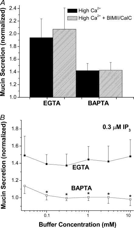
Figure 4. Lack of buffer effects on mucin secretion in SLO-permeabilized SPOC1 cells.
A, cells were exposed to high Ca2+ (3 μm), buffered by EGTA or BAPTA (0.1 μm Ca2+), ± the PKC inhibitors BIMII (1 μm) and calphostin C (1 μm; n = 4 SPOC1 passages). B, cells were exposed to IP3 (0.3 μm) and buffered by either EGTA or BAPTA (0.03–10 mm) at a constant 0.1 μm Ca2+ (n = 4 SPOC1 passages).

