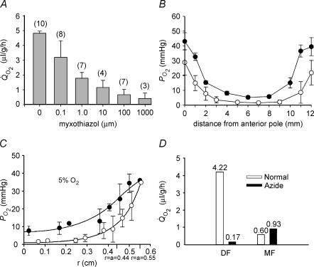
Figure 4. The effect of mitochondrial inhibitors on lens Q̇O2, PO2 profiles, and calculated oxygen consumption time constants.
A, lenses were pre-equilibrated in 5% oxygen for 4 h at 37°C. Lens Q̇O2 was inhibited by myxothiazol in a dose-dependent fashion. B, effect of myxothiazol on bovine lens PO2 profiles in vitro. Lenses were equilibrated in 5% O2 for 4 h at 37°C in the presence of 10 μm myxothiazol (•, n = 4). Compared to control lenses (○), myxothiazol treated lenses have a higher PO2 throughout the tissue. The PO2 gradient in the outer 2 mm of tissue is less steep in myxothiazol-treated lenses than controls. Note that, in each case, all data points are contained within the lens (the extreme data points nominally at 0 and 12 mm are in fact about 100 μm into the lens, explaining why the PO2 values are less than the values in the superfusion solution). C, incubation of lenses in 5 mm azide (•) also produces a generalized increase in PO2 and a shallower gradient in the superficial cell layers compared to untreated samples (○). The best fit of the diffusion–consumption model indicates that azide treatment causes a significant decrease in the rate of oxygen consumption in the outer shell of mitochondria-rich DF. D, the amount of oxygen consumed by the DF and MF cell layers in the presence or absence of azide was calculated using eqn (10) (Appendix). Note that, in addition to the time constants for oxygen consumption in DF and MF cells, the computed values also reflect the oxygen concentration in each region and the relative volumes of the two zones (see text for details). In the presence of azide, oxygen consumption by DF cells is dramatically reduced. In contrast, azide has little effect on oxygen consumption by the MF cells.

