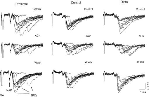
Figure 2.
Latencies of quantal release in proximal, central and distal regions of the synapse before (Control), in the presence of 5 × 10⁻⁴ m acetylcholine (ACh) and 60 min after washout (Wash). Recordings (9–11) were superimposed, showing extracellularly recorded presynaptic nerve action potentials (NAPs), individual endplate currents (EPCs) and stimulus artifacts (SA). The time intervals between the peak of the inward presynaptic Na+ currents of the nerve spike (downward deflection) and the times at which the rising phases of each EPC reached 20% of maximum was defined as the release latency. Note the most pronounced increase in latency dispersion in the distal region induced by ACh.

