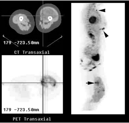
Figure 1.
Combined PET/CT imaging of a large soft tissue mass in the left thigh demonstrated marked heterogeneity of FDG uptake in the mass with small foci of very intense uptake superimposed on an overall pattern of only mildly to moderately increased activity. The reference transaxial CT and PET images correspond to the focus of highest FDG uptake seen on the right lateral maximum intensity projection images (horizontal arrow). Biopsy would normally have been directed at this site. However, multiple cutaneous metastases were identified (arrow heads). Note that the intensity of uptake at metastatic sites is similar to the intense primary tumoral uptake sites, consistent with the notion that the most metabolically active tumour sites are also the most aggressive and determine prognosis.

