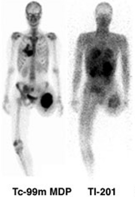
Figure 2.
99mTc MDP bone (left) and 201Tl (right) scans of a patient with recurrence of osteosarcoma in the left thigh and metastases to lung and bone are demonstrated. Although 201Tl demonstrates the peripheral and lung lesions quite well, the contrast is less than that observed on bone scanning, potentially limiting its sensitivity for other metastatic sites. The high uptake in abdominal and pelvic structures obscure the iliac and sacral metastases that are clearly seen on bone scan.

