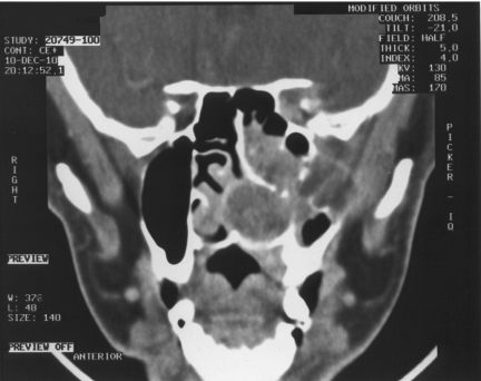
Figure 6.
Coronal CT post-contrast same patient as Fig. 5 with ACC. Demonstrating tumour throughout the left nasal cavity with bony destruction of the left maxillary sinus, loss of fat in the left infratemporal fossa and extension in the left orbital apex along the V2 division.

