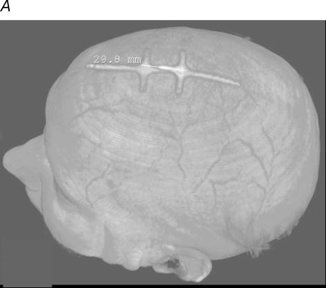
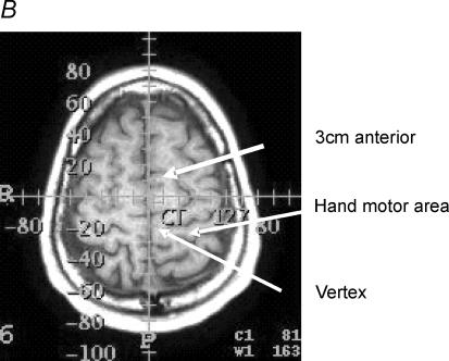
Figure 5. Magnetic resonance imaging.
Anatomical location of the primary motor cortex and supplementary motor area relative to the vertex and the 3 cm anterior position in one subject. A, markers used to identify the two positions. B shows that these markers were situated at −20 and +10 mm on an anterior–posterior axis and overlay the primary motor cortex (PMC) and supplementary motor area (SMA), respectively, which were identified according to Tallairach's handbook.


