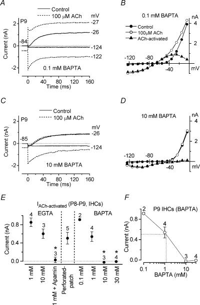
Figure 7. Effect of BAPTA on the ACh-activated SK current in immature IHCs.
Membrane currents (A and C) and steady-state I–V curves (B and D) recorded in P9 apical IHCs before and during superfusion of 100 μm ACh when 0.1 mm (A and B) and 10 mm (C and D) BAPTA were used in the intracellular solution instead of 1 mm EGTA. 0.1 mm BAPTA: Cm 9.5 pF; Rs 1.5 MΩ; gleak 1.0 nS. 10 mm BAPTA: Cm 9.6 pF; Rs 0.9 MΩ; gleak 3.0 nS. E, steady-state amplitude (at 160 ms) measured near −26 mV for the current during the superfusion of 100 μm ACh minus the control current using different intracellular Ca2+ buffers and using perforated-patch recording. F, size of the ACh-activated outward K+ current as a function of BAPTA concentration (data from E). The equivalent BAPTA concentration for the endogenous buffer is estimated by the dotted lines to be about 1 mm. All IHCs were from P8–P9 mice.

