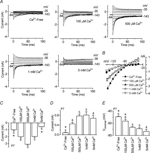
Figure 8. Extracellular Ca2+ both potentiates and blocks the ACh-activated current in immature IHCs.
A, isolated ACh-activated currents (P7, apical IHCs) obtained by subtracting the control currents from the currents in the presence of 100 μm ACh when a Ca2+-free solution or solutions containing different Ca2+ concentrations were superfused. All recordings were obtained using 0.9 mm extracellular Mg2+. Currents were recorded in response to a series of voltage steps from −144 mV to more positive potentials in 10 mV nominal increments (160 ms in duration) from the holding potential of −84 mV. Dotted lines indicate zero current. Ca2+-free (Ihold −145 pA) and 100 μm Ca2+ (Ihold−246 pA): Cm 8.6 pF; Rs 1.7 MΩ; gleak 2.8 nS. 500 μm Ca2+ (Ihold −459 pA) and 1 mm Ca2+ (Ihold −316 pA): Cm 6.8 pF; Rs 1.5 MΩ; gleak 1.2 nS. 5 mm Ca2+ (Ihold −71 pA): Cm 7.9 pF; Rs 2.0 MΩ; gleak 1.5 nS. B, average instantaneous I–V curves for the ACh-activated currents in the presence of different Ca2+ concentrations including those in A (numbers of cells as in C–E). C, size of instantaneous inward currents measured near −144 mV. Note that the size of the currents in both the Ca2+-free solution and the solution containing 5 mm Ca2+ was significantly smaller than that obtained in the presence of intermediate Ca2+ concentrations. D, size of instantaneous outward current measured around −35 mV. E, reversal potential of the instantaneous ACh-activated current as a function of extracellular Ca2+ concentration. Numbers of cells are shown above each column.

