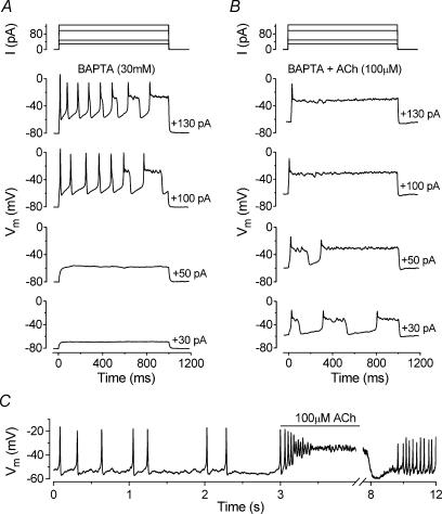
Figure 10. Effects of blocking the SK current on ACh-induced voltage responses in immature IHCs.
A and B, voltage responses from a P6 apical IHC before and during superfusion of 100 μm ACh, respectively. The intracellular solution contained 30 mm BAPTA to prevent the activation of the SK current. Current steps were applied from the resting potential in 10 pA increments between 0 and +130 pA and for clarity only a few voltage responses are shown. Superfusion of ACh resulted in resting membrane potential depolarization from around −80 mV (A) to −60 mV (B). Cm 9.0 pF; Rs 7.1 MΩ. C, continuous voltage recording obtained by applying a 10 pA depolarizing current injection to an apical P5 IHC. Before the recording shown the cell was superfused with 300 nm apamin. The line above the voltage recording indicates the period of ACh application. Note the compressed time scale of the abscissa after the interruption. Vm −59 mV; Cm 7.1 pF; Rs 5.2 MΩ; gleak 3.7 nS. All recordings at body temperature.

