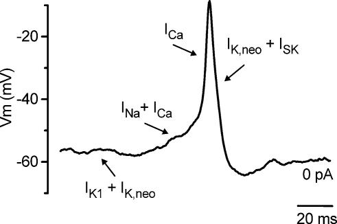
Figure 11. Role of different membrane currents in IHC action potential activity.
Spontaneous action potential recorded from an apical coil IHC (P3). The different basolateral currents expressed in immature IHCs exert distinct roles in different phases of the action potentials, as indicated by the arrows. See discussion for details.

