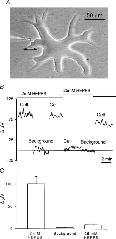
Figure 1. Self-referencing recordings from isolated skate horizontal cells using a H+-selective microelectrode.
A, an isolated cell obtained via enzymatic dissociation from the retina of the skate. The large size and thick cell soma permit ready identification of this cell as an external horizontal cell (Malchow et al. 1990). To the left is shown a H+-selective microelectrode positioned in the ‘near pole’ recording position. Differential recordings were made by laterally translating the electrode to a position 50 μm distant from the cell and computing the difference in signal between the near and far poles of the recording. The double arrow indicates the movement from near to far positions. B, a self-referencing differential recording from a single horizontal cell. The differential signal with the cell bathed in a skate Ringer solution containing 2 mm of the pH buffer Hepes was approximately 85 μV. Moving the electrode 250 μm (Background) resulted in a differential response close to 0 μV. Moving the electrode back to its original position restored the 85 μV signal. The solution was then replaced by one containing 25 mm Hepes; recording at the same location resulted now in a much smaller signal. This signal was still larger than that obtained at a background location in the same 25 mm Hepes solution. Replacing the solution with one containing 2 mm Hepes brought the signal near its original amplitude. C, average results from eight cells; recordings were first made with the cell bathed in 2 mm Hepes and the electrode 1 μm from the cell membrane. Differential recordings were then obtained at a position at least 250 μm from the cell (Background). The solution around the cell was then changed to one containing 25 mm Hepes and the response recorded at the original position of the electrode near the cell.

