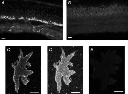
Figure 11. Immunocytochemical localization of the plasmalemma Ca2+–H+-ATPase protein in isolated retina and isolated horizontal cells.
A, staining pattern of fluorescence in a transverse section through the skate retina. Intense staining is distinctly visible at the level of the outer synaptic layer; lighter staining is evident within the inner synaptic layer. B, control section showing florescence seen in the absence of the primary antibody. C, confocal optical section through a single external horizontal cell stained for the plasmalemma Ca2+–H+-ATPase. Note the bright staining observed along the edges of the cell membrane. D, optical stack showing complete staining pattern for the same horizontal cell as shown in C. Note the intense staining on the cell membrane surface. E, control photomicrograph of a horizontal cell in which the primary antibody was omitted during the staining procedure. Scale bars are 10 μm for A and B, 35 μm for C–E.

