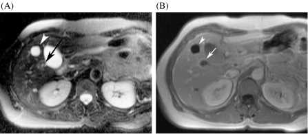
Figure 4.
Small metastasis and cyst: differentiation with T2-weighted TSE and non-specific gadolinium chelates. (A) The T2-weighted TSE image shows a small cyst, which is very bright (arrowhead). There is a second lesion, which is moderately hyperintense (arrow). (B) The gadolinium-enhanced T1-weighted GRE image shows lack of enhancement of the cyst (arrowhead). The other lesion displays a ring enhancement, which is suggestive of metastasis (arrow).

