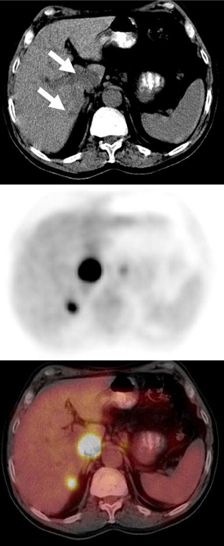
Figure 4.
A staging FDG-PET/CT scan in a patient with esophageal cancer demonstrated intense FDG avidity in two hypodense liver lesions (large lesion in the caudate lobe and a smaller lesion in the medial aspect of the posterior segment of the right hepatic lobe) consistent with liver metastases. The CT scan provided accurate anatomic localization of the abnormal FDG activity to the liver lesions, clearly excluding the right adrenal gland or paraaortic lymph nodes as sites of metastatic disease.

