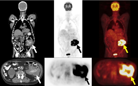
Figure 9.
A 78-year-old man with right temple melanoma (Breslow depth 2.28 mm), after wide local excision 6 years previously, presented with anemia and a left upper quadrant mass. A restaging FDG-PET/CT study demonstrated an intensely FDG avid mass associated with thickened jejunal loops of bowel (arrows). Biopsy and subsequent surgery diagnosed metastatic melanoma involving the proximal jejunum.

