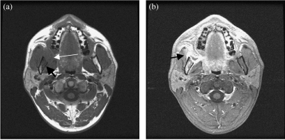
Figure 2.
(a) Axial T1-weighted MRI shows a right intermediate signal intensity retromolar trigone carcinoma (thin arrow). There is adjacent mandibular marrow involvement (thick arrow). (b) Axial contrast enhanced MRI shows tumour enhancement. Note the marrow enhancement and involvement of the right masseter muscle (arrow).

