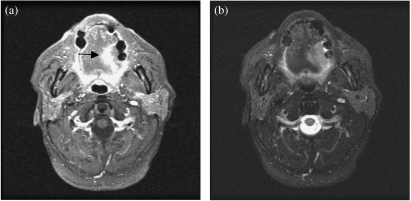
Figure 3.
(a) Axial contrast enhanced MRI shows an enhancing tumour on the lateral aspect of the left hemitongue (arrow). (b) Axial T2-weighted MRI shows a high signal intensity left tongue carcinoma. Fat saturated T2-weighted MRI is particularly useful in separating the lesion from normal tongue tissue.

