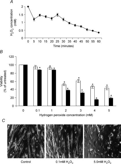
Figure 2. The effect of H2O2 treatment on the viability of C2C12 myotubes.
A, change in absolute concentration of H2O2 with time during incubation in Dulbecco's phosphate-buffered saline. The initial concentration of H2O2 was 2 mm. B, viability of 5-day-old cultured myotubes measured at either 4 (□) or 12 (▪) hours following exposure to different concentrations of H2O2 for 30 min *P < 0.05 compared to untreated cells; n = 12. C, light microscopy of control untreated 5-day-old myotubes and myotubes at 4 h following treatment with 0.1 or 2 mm H2O2 for 30 min.

