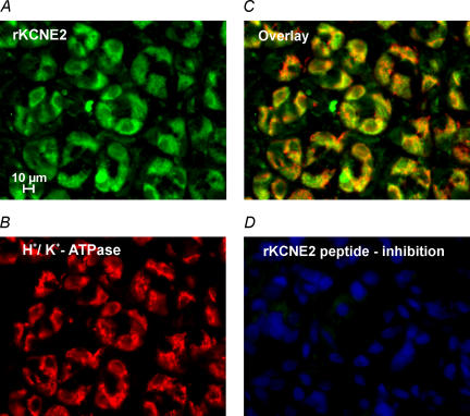
Figure 3. KCNE2 in non-stimulated rat gastric mucosa.
A, epifluorescence images of rat oxyntic mucosa stained with polyclonal, affinity-purified KCNE2 antibody. B, H+,K+-ATPase labelling with monoclonal antibody. C, overlay of KCNE2 and H+,K+-ATPase staining shows partial co-localization in parietal cells as described for KCNQ1 and H+,K+-ATPase. D, no staining was observed with the KCNE2 antibody after absorption to the peptide used for immunization; nuclear staining with HOE33342 is visible.

