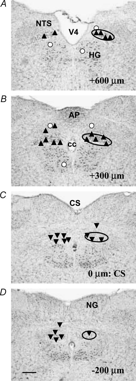
Figure 2. Summary map showing the location of the sites of microinjection of PrRP into the DMV at levels corresponding to the rostral pole of the nucleus (A), the area postrema (B), CS (C) and caudal pole of the DMV (D).
The approximate location of the DMV is outlined by the solid oval. For the purposes of this study, the portion of the DMV extending from the CS to +0.2 mm rostral of the anteriormost border of the area postrema was taken to define the rostral DMV (A and B) and the region extending caudally from the CS (C and D) was defined as the caudal DMV. Numbers in lower right of panels indicate rostral–caudal distance measured relative to the CS, with the caudalmost tip of the area postrema set as 0 μm. Upward triangles indicate sites where microinjection of PrRP increased IGP, whereas downward triangles indicate sites where IGP was decreased in response to PrRP. Open circles indicate sites where administration of the peptide did not evoke changes in IGP. Locations of various NTS subnuclei are not shown for purposes of clarity. Abbreviations: AP, area postrema; cc, central canal; CS, calamus scriptorius; HG, hypoglossal nucleus; NG, nucleus gracilis; NTS, nucleus tractus solitarius; V4, fourth ventricle. Scale bar=300 μm.

