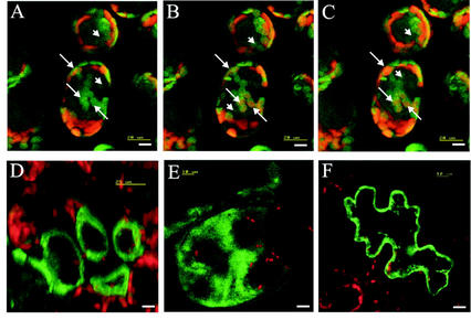
Figure 4.
Transient expression of PLA-GFP fusion proteins in tobacco leaf cells as visualized by confocal laser scanning microscopy. A through C, Three consecutive optical sections, moving from the epidermal top side of palisade cells toward the spongy parenchyma side containing, expressed PLA-I-GFP fusion protein. Arrows, Localization of PLA-I-GFP fusion protein in an individual chloroplast in consecutive frames. Arrowheads, Localization of PLA-I-GFP fusion protein in the cytosol. D, Single optical section of palisade parenchyma cells expressing the PLA-IIA-GFP fusion protein. E, Summarized image showing the expressed PLA-IVA-GFP-fusion protein calculated from 16 consecutively taken single optical sections (in z-direction) through a large cell. F, Single optical section of an epidermal cell expressing the PLA-IVC-GFP fusion protein. Bar = 10 μm.

