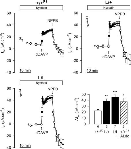
Figure 7. Effects of basolateral membrane permeablilization on dDAVP-stimulated Isc.
Isc was measured in sets of confluent cultures of +/+(L), L/+ and L/L CCD cells grown on filters after the basolateral membrane had been permeabilized by adding nystatin (360 μg ml−1) to the basal side of the filter, and a Cl− gradient (basal medium: 149 mm; apical medium: 14.9 mm) imposed as described in the Methods. Nystatin (^) was added to the basal medium for 20 min. The cells were then sequentially incubated with basal dDAVP (10−8m, •) for 10 min, and then with apical NPPB (10−4m, δ) for a further 10 min. Similar Isc recordings were performed on +/+(L) CCD preincubated with 10−6m aldosterone for 4 h (+/+(L)+Aldo) before permeabilization of the basolateral membrane (hatched bar). Bars represent the relative δIsc increase caused by dDAVP. Values are means ± s.e.m. from five to seven separate experiments. *P < 0.05, **P < 0.01, ***P < 0.001versus untreated +/+(L) values.

