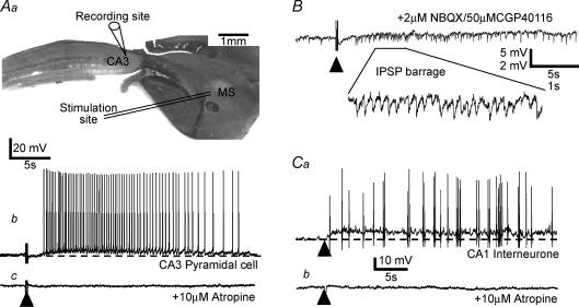
Figure 1. Activation of septo-hippocampal afferents excites hippocampal pyramidal cells and interneurones.
Aa, diagram of septo-hippocampal slice showing relative position of stimulating (within medial septum, MS) and recording electrode (CA3 pyramidal cell). Ab, intracellular recording from a CA3 pyramidal cell reveals an isolated slow depolarizing response to electrical stimulation (indicated by ▴) within the septal nucleus in the presence of a cocktail of AMPA/kainate, NMDA, GABAA and GABAB receptor antagonists (4 μm NBQX, 50 μm CGP40116, 50 μm picrotoxin and 1 μm CGP55845A, respectively). Ac, the action potential discharge and underlying slow depolarizing waveform were abolished upon application of the mAChR antagonist atropine (10 μm). B, example of a similar experiment in which only the AMPA/kainate and NMDA receptor antagonists were present to block glutamatergic EPSPs. The presence of a barrage of IPSPs (shown also in expanded inset) following afferent stimulation suggests a direct cholinergic excitation of presynaptic GABAergic interneurones. Subsequent application of 10 μm atropine abolished IPSP trains in this cell following afferent stimulation (data not shown). Ca, recording from a putative fast-spiking interneurone within area CA1 in which a similar slow depolarizing response is evoked following stimulation of cholinergic afferents. Cb, as with the pyramidal cell response, the slow depolarizing potential is completely abolished upon subsequent coapplication of the mAChR antagonist atropine (10 μm). Detail of evoked cholinergic EPSP methodology given in Morton & Davies (1997).

