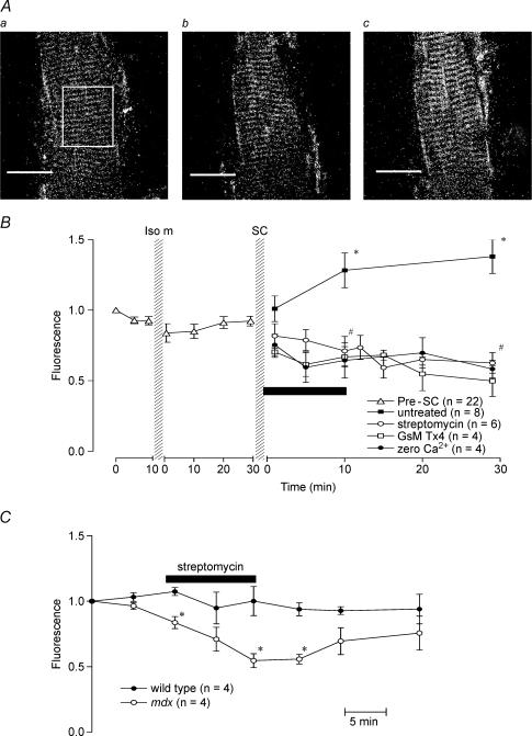
Figure 1. [Ca2+]i signals from mdx muscle fibres.
A, confocal images of fluo-3 fluorescence from one mdx muscle fibre under control conditions (a), following isometric contractions (b) and 30 min after stretched contractions (c). Fluorescence intensity was measured by outlining a region of the muscle fibre with a rectangle as indicated in a. Scale bars represent 50 μm. B, the effect of stretch-activated channel blockers and zero extracellular Ca2+ on resting [Ca2+]i following stretched contractions (SC) in mdx muscle fibres. The fluorescence intensity was normalized to the starting point of each experiment. Following stretched contractions in the untreated fibres, [Ca2+]i-dependent fluorescence was significantly higher than before the stretched contractions (*). Application of stretch-activated channel blockers, streptomycin or GsMTx4, or zero Ca2+ solution for 10 min following stretched contractions (as indicated by the horizontal bar) led to a reduction in [Ca2+]i. #Significant difference between the treated and untreated experiments. Bars represent s.e.m. C, effect of streptomycin on resting [Ca2+]i in wild-type and mdx muscle fibres. Muscle fibres were exposed to 200 μm streptomcyin for 10 min as indicated by the bar. *Significant difference between wild-type and mdx muscle fibres. Bars represent s.e.m.

