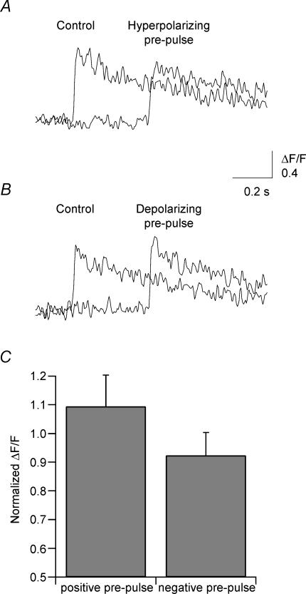
Figure 8. Modulation of dendritic Ca2+ transients by the waveform of somatic APs.
A, fluorescence traces recorded from the dendrite of bitufted interneurone using two-photon scanning laser microscope. The left trace represents Ca2+ transient in response to a b-AP generated by current injection into the interneurone soma. The amplitude of Ca2+ transient decreased (right trace) when a 400-ms prepulse of hyperpolarizing current (deflecting the membrane potential by −10 mV) was injected into the soma. B, the amplitude of Ca2+ transient became larger when the same experiment was repeated with a current prepulse depolarizing the membrane by 10 mV (right trace). D, summary of four experiments showing the change in ΔF/F, normalized to control, following a positive prepulse and a negative prepulse. Error bars are s.e.m.

