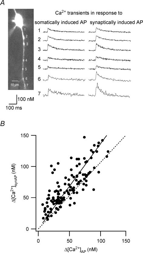
Figure 10. Synaptically initiated and somatically initiated APs evoke different dendritic Ca2+ transients.
A, fluorescence Ca2+ imaging of a bitufted interneurone synaptically connected to a neighbouring pyramidal neurone (not shown); rectangles mark dendritic regions selected for imaging. Ca2+ transients caused in response to a single AP evoked by either somatic current injection into the interneurone (left) or unitary synaptic stimulation (right). Six sweeps were averaged. B, relationship between Ca2+ transients evoked by somatically initiated and synaptically initiated b-APs. Data from 134 dendritic regions of 23 interneurones. Data fitted by linear regression with a slope of 1.37 ± 0.03. Ca2+ amplitude was ∼2.5-fold larger when corrected for the presence of exogenous Ca2+ buffer (Fura-2) (Kaiser et al. 2001).

