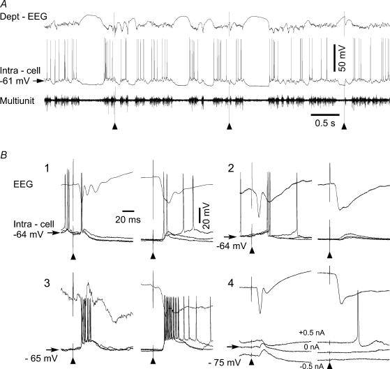
Figure 1. Modulation of cortical somatosensory evoked potential and intracellular activities during slow oscillation.
All recordings were obtained from the focus of responses to the electrical stimulation of the right forelimb median nerve in the left somatosensory cortex. A, an example of the focal field, intracellular and multiunit recordings during a session of peripheral stimulation (▴). The first and the second stimuli were applied during active network states and the third stimulus was delivered during the silent network state. B, averaged field potential responses and superimposition of three intracellular single traces selected during active (left side) and silent (right side) network states. Note the multiple components of field responses during active cortical states and their modification during silent states. 1, 2 and 3 are the three types of initial excitatory responses; 4 is the initial inhibitory response. Note the increased latency of excitatory responses that occurred during EEG depth-positive wave and the absence of inhibitory response during the silent network state.

