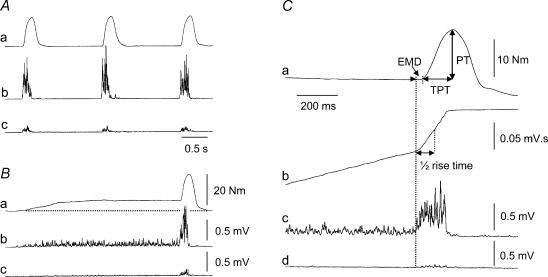
Figure 1. Contraction protocol and measurements.
Torque (a) and rectified surface EMG from the tibialis anterior (b) and soleus (c) during ballistic contractions of the ankle dorsiflexors performed from a resting state (A) or superimposed on a sustained muscle activation at 25% of MVC (B) in one subject. C, illustration of the different measurements on the torque (a) and EMG signals of the tibialis anterior (b and c) and soleus (d) during a ballistic contraction with pre-activation. The peak torque (PT) achieved during the ballistic contraction was measured as the difference between PT and baseline torque, which was zero without pre-activation and ∼25% PT with pre-activation. The time to peak torque (TPT) of the ballistic contraction was determined from its onset to the peak torque. The electromechanical delay (EMD) was defined as the time lag between the onset of the agonist EMG burst and of the torque. The time to reach 50% of the maximal EMG (½ rise time) during the ballistic burst was measured from the integrated EMG signal (b).

