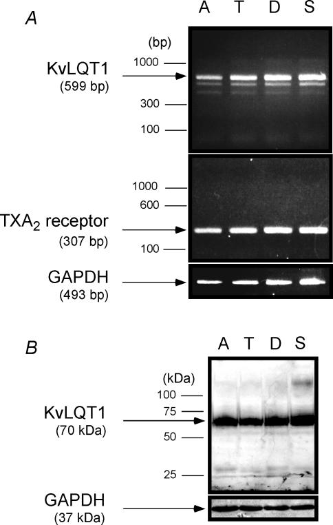
Figure 5. Expressions of TXA2 receptor and KvLQT1 in the human colonic mucosa.
A, gel analysis of RT-PCR products from human colonic mucosa isolated from ascending colon (A), transverse colon (T), descending colon (D) and sigmoid colon (S). Each specimen was derived from a different patient. A single band of 599 bp (KvLQT1; upper panel) and 307 bp (TXA2 receptor; middle panel) were detected by ethidium bromide staining. Amplification of GAPDH (493 bp) was used for control (lower panel). B, Western blotting for detecting the KvLQT1 protein. The blotting was performed with 80 μg of membrane protein from the same specimens as used in A. Expression of the KvLQT1 protein (70 kDa) is observed in all lanes (upper panel). As a control, expression of GAPDH protein (37 kDa) was examined (lower panel).

