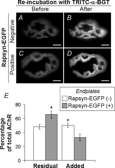
Figure 9. Loss and replacement of AChRs from endplates.
Four days after electroporation of rapsyn–EGFP plasmid, the surface of the TA muscle was again exposed, and AChRs were saturated with TRITC-α-BGT. A and C, on day 8 after electroporation (4 days after TRITC-α-BGT incubation), endplates negative (A) and positive (C) for rapsyn–EGFP were imaged to reveal AChRs retained from the time of TRITC-α-BGT-labelling 4 days earlier (Residual AChR). The same endplates were imaged again after re-labelling with TRITC-α-BGT to reveal total AChR (B and D). E, quantitative analysis of TRITC-α-BGT fluorescence at endplates (as shown in A–D). Residual AChR and added AChR (additional AChR revealed by re-labelling with TRITC-α-BGT) were normalized to total TRITC-α-BGT intensity. Rapsyn–EGFP-positive endplates showed higher levels of residual AChR and lower levels of replacement (Added) AChRs compared with rapsyn–EGFP-negative endplates (*P < 0.05; n = 7 for each group). Scale bar, 10 μm.

