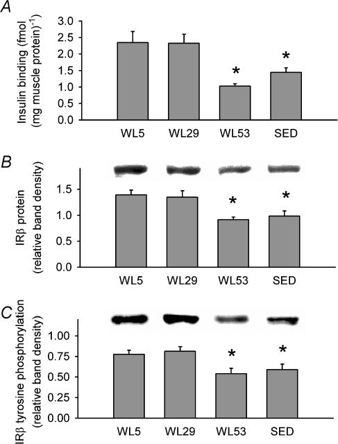
Figure 3. Effect of decreased physical activity on descriptive indices of the insulin receptor and its activation in epitrochlearis muscle.
A, submaximal insulin binding to the epitrochlearis muscle. B, IRβ protein levels relative to loading control (see Methods) with a representative immunoblot (above graph). C, tyrosine phosphorylation of IRβ in response to 40 min of submaximal insulin stimulation with a representative immunoblot (above graph). IRβ immunoprecipitates were subjected to immunoblotting for phosphotyrosine, then stripped and re-probed for IRβ (see Methods). Data were normalized to a loading control (see Methods) and are expressed relative to the normalized band intensity for IRβ protein present in the same lane. Columns are mean ± s.e.m.* Signficantly different (ANOVA, P≤ 0.05) from groups without an asterisk. n = 6–8 in each group.

