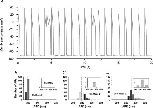
Figure 6. Early after-depolarizations (EADs) during β-AR stimulation.
A, membrane potential (millivolts) as a function of time (seconds) for the β-AR-stimulated model in response to 1 Hz pacing; action potential duration (APD) distribution in simulations of 150 APs with 0% (B), 15% (C), or 25% (D) of LCCs gating in mode 2 (10 ms bins) Reproduced from Tanskanen et al. (2005) with permission from the Biophysical Society.

