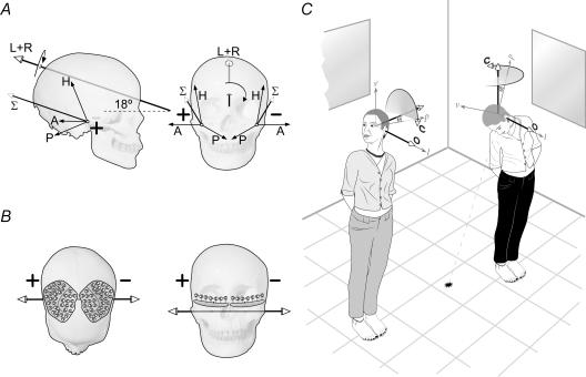
Figure 1. Predictions and methods.
A, predicted GVS-evoked signal from the semicircular canals. The anatomical orientation of the three canals and their receptor cells will produce specific rotational vectors from the horizontal (H), anterior (A) and posterior (P) canals. These are shown for bilateral bipolar GVS with anodal current (+) on the right and cathodal current (−) on the left. The vector sums from each side (Σ) add to create a final resultant (L + R). This GVS canal vector is directed backward and upwards by 18 deg from Reid's plane (dashed line). It is in the sagittal plane and will therefore change orientation with head pitch. B, the predicted GVS signal from the utricles relies on the imbalance between medially and laterally orientated hair cells. It is horizontal in the coronal plane and therefore does not change with head pitch. C, in this experiment, GVS is delivered with the head in two positions. Head-up has the canal vector (C) horizontal and head-down has it vertical, whereas the otolith vector (O) is the same in both positions. Arrows v, l and p indicate vertical, lateral and posterior in head coordinates. The plane lp is Reid's plane.

