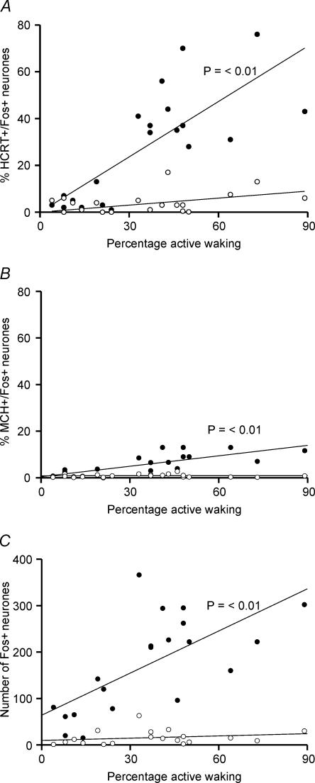
Figure 9. Regression functions and correlation between bicuculline-induced active waking and increase in Fos-IR.
Regression lines and scatter plots of the percentage of HCRT+/Fos+ (A), MCH+/Fos+ (B) and the number of single Fos+ neurones (C) in the ipsilateral side (•) and the contralateral side (○) versus percentage active wake time during the 2-h recording period before the rats were killed. The bicuculline-induced Fos-IR in a different neuronal population ipsilateral to the microdialysis probe was positively correlated with the amount of active waking. No significant correlation was found for neurones on the contralateral side. The active–wake and Fos-IR data were pooled from all rats used in this study for aCSF and bicuculline treatments.

