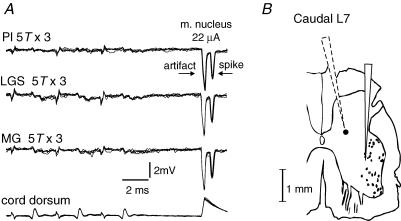
Figure 4. Examples of records from a candidate excitatory interneurone which is unresponsive to group I stimulation in the absence of locomotion.
A, overlayed extracellular recordings (n = 5) from an interneurone that was antidromically activated by stimuli applied in the ankle extensor motor nuclei (latency, 0.6 ms) but failed to respond to stimulation supramaximal for group I afferents (5T × 3, 300 Hz). Bottom trace is the cord dorsum recording of the afferent volleys. B, camera lucida reconstruction of the recording microelectrode track (dotted outline) indicating the estimated location (filled circle) of the interneurone illustrated in Figs 4A, 5, 6 and 8A. The solid outline to the right is the track of the tungsten stimulating electrode. The scale bar takes into account a 10% shrinkage due to mounting. This interneurone was located in the intermediate nucleus.

