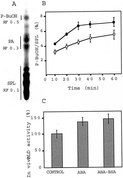
Figure 2.
In vivo PLD activity is stimulated by ABA in Arabidopsis suspension cells. A, Thin-layer chromatography separation of phospholipids after treatment with ABA-BSA (10−5 m equivalent ABA) and 0.5% (v/v) 1-BuOH after an 18-h 33P pulse. B, Time course of in vivo ABA-stimulated PLD activity. ○, Control; ●, ABA (10−5 M). Bars, sd, n = 3. C, Free (10−5 m) or conjugated (10−5 m equivalent ABA) ABA-stimulated PLD activity. PLD activity was measured after 30 min of treatment. SPL, Structural phospholipids. Bars, sd, n = 7.

