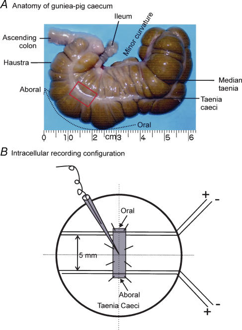
Figure 1. Anatomy of taenia caecum and intracellular recording configuration.
A, photograph of guinea-pig caecum. The taenia caecum is clearly visible as a distinct thickening of longitudinal muscle, with the aboral end being the portion closest to the ascending colon. The framed area represents the portion dissected free for intracellular recording. B, intracellular recording set up. Two pairs of silver wires were placed at the oral and aboral ends of the tissue 5 mm far from each other. Intracellular recording was made in the middle of two pairs of stimulating electrodes. In all figures except Fig. 9, recordings depict results obtained with oral NS.

